Which diseases can be diagnosed through a comprehensive MRI scan of the entire spine? This imaging procedure covers the cervical, thoracic, and lumbar regions, offering a detailed view of the spinal cord. Specifically, a cervical spine MRI scan is employed to assess pain, injuries, and numbness extending to the arm(s).
What is Whole Spine MRI Screening?
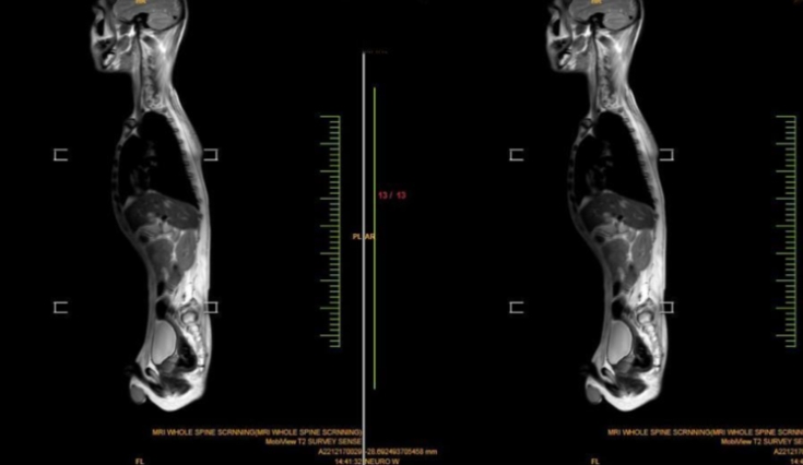
A whole spine MRI screening serves as a non-invasive diagnostic method, utilizing advanced equipment that generates magnetic fields and radio waves. This process yields a precise and clear depiction of the entire spine, encompassing the spinal cord, discs, muscles, and surrounding soft tissues.
What are some typical applications of this procedure? Magnetic Resonance Imaging (MRI) is employed for various purposes, including the evaluation or identification of:
Spinal anatomy and its alignment.
Birth defects affecting the vertebrae or spinal cord.
Traumatic injuries involving the bone, disc, ligament, or spinal cord.
Diseases affecting the discs and joints, which often contribute to severe lower back pain and sciatica (back pain radiating into the lower leg).
Compression or inflammation of the spinal cord and nerves.
Infections affecting the vertebrae, discs, spinal cord, or its coverings (meninges).
Tumors located in the vertebrae, spinal cord, nerves, or the surrounding soft tissues.
Spine MRI is utilized to assist in planning medical procedures like decompression of a pinched nerve, spinal fusion, or administering steroid injections. These injections, conducted under x-ray guidance, alleviate pain. Spine MRI also identifies other potential causes of back pain, such as compression fractures and bone swelling. Additionally, it is employed for monitoring changes in the spine post-surgery, such as scarring or infection.
Anatomy and Function of Your Spine
Also called the “backbone,” your spinal column is a complex group of bones that creates your body’s main support. The bones of the spine are called vertebrae. You have 33 of them stacked on top of each other, interlocking to form the column that surrounds your spinal cord. A healthy spine actually has a natural S-shaped curve.The five sections or segments of your spine are: cervical, thoracic, lumbar, sacral, and coccygeal.
Between each vertebra are tiny shock absorbers called intervertebral discs. Ligaments are soft tissues that hold the vertebrae together, and tendons connect them to muscles. Your spine works with your nervous and musculoskeletal systems to make sure you can sit, twist, bend, and walk.
How Long Will Whole Spine MRI Screening Take?
Unlike X-rays, which use harmful ionizing radiation, magnetic resonance imaging uses a magnetic field and radio waves to take detailed images of your spine and nearby tissues.
An MRI exam is useful in evaluating the medical conditions impacting the soft tissue structures of the back, including the position of the vertebrae that make up your spinal column. It may spot abnormalities indicative of infection, nerve and disc problems, arthritis, blood vessel problems, and spinal tumors.
What Information Can a MRI Cervical Spine Screening show?
The cervical spine, also known as the C-spine, comprises the initial seven vertebrae in your spinal column. A cervical spine MRI examines the neck region from the base of your head to the commencement of the thoracic or mid-back area. It encompasses various structures such as the thyroid gland, throat, larynx, neck muscles, ligaments, tendons, and other soft tissues.
Several types of abnormalities that a cervical spine MRI procedure may identify include:
- Congenital birth defects
- Scarring or injury
- Irregularities in the position of vertebrae or in the curvature of the spine
- Pinched nerves
- Cancer tumors in the cervical spine
- Thyroid gland tumors
- Spinal stenosis, characterized by a narrowing of the spinal column
- Cysts
What Insights Can MRI Thoracic Spine Screening Provide?
The thoracic segment comprises 12 larger vertebrae compared to the cervical vertebrae and is situated in the upper or middle back. This region includes muscles, thoracic ligaments, tendons, and intervertebral discs. The thoracic spine also consists of 12 sets of ribs and structures contributing to the thoracic cavity, with joints attached through another type of soft tissue known as cartilage.
Various abnormalities that may be observed on a thoracic spine MRI are:
- Tumors in the spinal canal
- Tumors of the spinal cord
- Bulging spinal discs
- Compression fractures
- Disc degeneration
- Cysts
- Injuries to nerves, muscles, ligaments, and tendons
- Injuries to the rib cage and costochondral separation (where the rib pulls away from the cartilage)
- Joint inflammation
- Abscesses and other signs of infection
What Information Can You Obtain From MRI Lumbar Spine Screening?
The lumbar spine, constituting the lower back, typically includes the sacral and coccygeal regions as well. Comprising five larger vertebrae than those in the thoracic region, the lumbar spine connects the thorax to the pelvis and sacrum. It involves large muscles for bending, lifting, and carrying heavy loads, along with a complex network of nerves and blood vessels.
During a lumbar MRI, various abnormalities that may manifest include:
- Bulging or herniated disc
- Degenerative disc disease
- Disc height loss
- Vertebral malformation
- Muscle, tendon, and ligament injury
- Abnormal nerve roots, including nerve injury
- Spine infections
- Sclerosis
- Neuromuscular diseases
- Tumors
- Aneurysms and other blood vessel disorders
- Cysts
Whole Spine MRI Screening Technology in Forethought
Forethought Professional Version II utilizes whole spine MRI screening technology to provide patients with a comprehensive, radiation-free testing solution for precise tracking, follow-up and treatment adjustments of cervical spine conditions.
Innovative Intelligent Light Sensing Technology
Forethought Professional Version II introduces advanced intelligent light sensing technology, which realizes real-time monitoring of the cervical spine condition through the use of MEMS sensors to dynamically capture minute angular velocity changes.
Accurate Terrain Scanning Technology
Depending on the operator’s speed, Forethought Professional Version II utilizes precise topographic scanning technology to collect optimal topographic data, providing an accurate scanning base for cervical spine MRI.
Accurate Topographic Scanning Technology with Multi-level, Multi-spatial Information
Forethought Professional Version II features precise topographic scanning technology with multi-level, multi-spatial information that is integrated and optimized for processing, providing physicians with comprehensive, complementary data to aid in accurate diagnosis and treatment planning.
https://www.forethoughtmed.com/what-is-Included-in-whole-spine-mri-screening.html